Main Types of Microscopes
The table below describes the main types of microscopes within the optical, electron, and scanning probe categories.
Optical microscope
| Type | Description |
|---|---|
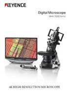
| Digital microscope | A microscope that uses a camera and magnified optics. It enables to output a live image to a monitor. |
| Binocular stereoscopic microscope | A microscope that allows easy observation of 3D objects at low magnification. |
| Brightfield microscope | A typical microscope that uses transmitted light to observe targets at high magnification. |
| Polarizing microscope | A microscope that uses different light transmission characteristics of materials, such as crystalline structures, to produce an image. |
| Phase contrast microscope | A microscope that visualizes minute surface irregularities by using light interference. It is commonly used to observe living cells without staining them. What is a phase contrast microscope? With a conventional biological microscope, it is difficult to observe colourless, transparent cells while they are alive. A phase contrast microscope makes it possible by utilizing two characteristics of light, diffraction and interference, to visualize specimens based on brightness differences (contrast).
|
| Differential interference contrast microscope | This microscope, similar to the phase contrast, is used to observe minute surface irregularities but at a higher resolution. However, the use of polarized light limits the variety of observable specimen containers. |
| Laser microscope (Laser scanning confocal microscope) |
This microscope uses laser beams for clear observation of thick samples with different focal distances. |
| Multiphoton excitation microscope | The use of multiple excitation lasers reduces damage to cells and allows high-resolution observation of deep areas. This type of microscope is used to observe nerve cells and blood flow in the brain. |
| Structured illumination microscope | A high-resolution microscope with advanced technology to overcome limited resolution found in optical microscopes that is caused by the diffraction of light. What is a structured illumination microscope? A type of high-resolution microscope based on technology that has overcome the limited resolution of optical microscopes caused by the diffraction limit of light.
|
Electron microscope
| Type | Description |
|---|---|
| Transmission electron microscope (TEM), scanning electron microscope (SEM), etc. | These microscopes emit electron beams, not light beams, toward targets to magnify them. |
Scanning probe microscope (SPM)
| Type | Description |
|---|---|
| Atomic force microscope (AFM), scanning near-field optical microscope (SNOM), etc. | This microscope scans the surface of samples with a probe and this interaction is used to measure fine surface shapes or properties. |
Others
| Type | Description |
|---|---|
| X-ray microscope, ultrasonic microscope, etc. | - |
In addition to the above categories, optical microscopes can be classified as follows:
Classification by application
| Biological microscope | With a magnification ranging from 50x to 1,500x, this microscope uses sliced samples that are fixed onto slides for observation. |
|---|---|
| (Binocular) stereoscopic microscope | The binocular system allows 3D observation of samples, such as insects or minerals, in their natural state without the need to be sliced. The magnification ranges from 10x to 50x. |
Classification by structure
| Upright microscope | Observes targets from above. This type of microscope is used to observe specimens on slides. |
|---|---|
| Inverted microscope | Observes targets from below. This microscope is used to observe, for example, cells soaked with culture in a dish. |
Magnified Observation and Instruments
A microscope is an optical instrument used to view small objects by enlarging them with two convex lenses. Optical microscopes, used for research, illuminate samples with visible or ultraviolet light. Depending on its structure, a biological microscope is categorized as an upright or inverted with a magnification ranging from 10x to 1500x.
Different types of microscopes are used based on the desired level of magnification. Magnifying glasses or loupes are used for quick inspection with a low magnification; binocular microscopes are used to observe from 10x to 50x, and upright/inverted microscopes are used to observe from 50x to 1500x.
Viewable objects by magnification
| Magnification | Instrument | Example |
|---|---|---|
| 1x | Naked eye | Hair (approx. 0.1 mm) |
| Approx. 2x to 5x | Magnifying glass | Plant or insect |
| Approx. 10x to 20x | Stereoscopic microscope | Water fleas and other microorganisms |
| Approx. 50x | Upright/inverted microscope | Insect's compound eye |
| Approx. 100x | Upright/inverted microscope | Paramecium |
| Approx. 200x | Upright/inverted microscope | Pollen |
| Approx. 400x | Upright/inverted microscope | Euglena |
| Approx. 800x to 1,500x | Upright/inverted microscope | Cell or chromosome (approx. 0.2µm) |
| Approx. 2,000x to 1,000,000x | Electron microscope | Objects from 1μm to 0.1 nm such as a DNA (2 nm) |
Trivia: What is the reference for a magnification of 1x?
A magnification of 1x is based on the point where a nearby object can clearly be observed by the human eye. Because this distance is 250 mm (distance of distinct vision), the size that can be observed at this distance is specified as 1x.