Sophistication and Efficiency Improvement of Observation and Analysis in Forensics Such as Medical Jurisprudence
In detailed investigation of incidents or crimes and verification of evidence that determines guilt or innocence, it is very important to examine and inspect evidence collected at the scenes in a sophisticated, scientific, and precise manner. Therefore, collected evidence needs to be analysed from various perspectives such as medical jurisprudence, chemistry, and physics.
This section introduces application examples of our 4K digital microscope, which further improves the sophistication of observation and measurement in forensics.
- Examination Fields and Microscope Applications in Forensics
- Application Examples of Our 4K Digital Microscope in Forensics Including Medical Jurisprudence
- Features of the VHX Series 4K digital microscope
- High-magnification observation of hair
- Observation of lustrous evidence such as metal dental crowns
- Capturing of fingerprints with high-resolution 4K images
- High-magnification observation of fibres
- Image capturing of pollens in filters
- Handwriting analysis using high colour gradation images and colour map images
- A 4K Digital Microscope That Improves the Sophistication and Efficiency of Forensic Work Including Medical Jurisprudence
Examination Fields and Microscope Applications in Forensics
Forensics includes medical jurisprudence, chemistry, and physics. Specialised examination and inspection are performed in each field. Read on for an explanation of the representative fields in forensics and for overviews of them.
Representative fields in forensics
- Biological examination
- This field analyses, examines, and inspects biological samples—such as blood, bones, hair, and saliva—related to an incident or crime from the medicolegal perspective.
- Chemical identification
- This field chemically examines and inspects materials—such as coating and fibres left at scenes of incidents, oils collected at scenes of fires, and illegally dumped industrial waste—as well as inspects drugs and poisons related to illegal drug cases and poisoning death cases.
- Physical identification
- This field estimates the circumstances of accidents and identifies causes of fire cases and traffic accidents by performing on-site investigations and reproductive experiments. This field also analyses security footage, images, and recorded sounds and voices and also examines and inspects materials such as guns and bullets.
- Document examination
- This field examines and inspects documents. For example, identification of handwriting and seals on documents, such as contracts and letters; identification of printed fraudulent materials, such as paper currencies, cash vouchers, and driver licences; identification of printers used for threatening letters; and detection of signs of doctoring. When psychological examination and inspection are required in addition to document examination, a polygraph is used.
Microscope applications in forensics
In forensics, components of evidence, blood, DNA, and other related materials are analysed. Additionally, it is important to use microscopes to capture, observe, and analyse the appearance in a sophisticated and non-destructive manner. Some concrete examples are given below.
- Fingerprint identification
- Handwriting identification
- Fibre identification
- Adhering substance identification
- Image capturing of skin, hair, and dental features
- Identification of plankton in fluids left in airways
- Image capturing of rifling marks
- Image capturing of signs of cutting and melting
- Identification of printed fraudulent materials
Application Examples of Our 4K Digital Microscope in Forensics Including Medical Jurisprudence
No mistake is allowed in forensic examination because the results can influence the lives of people involved in crimes or incidents. It is also important for forensics to capture and submit images that can be very clear evidence. To use microscopes for forensic observation and analysis, the equipment needs to have high performance and high reliability and users need to have a high level of technical skill and experience in using this equipment.
For over 30 years, KEYENCE have developed microscopes that are used by companies and research institutes all around the world, thoroughly pursuing usability as well as reliability by achieving higher performance and functionality.
The result of the latest technologies and observation and analysis knowledge is the VHX Series 4K digital microscope.
This microscope uses a cutting-edge optical system, a 4K CMOS image sensor, various image processing functions, and an observation system under motorised control. These features allow for sophisticated and precise observation and analysis with simple operations. Read on for an introduction to the VHX Series’ features and usage examples applicable to forensics.
Features of the VHX Series 4K digital microscope
- A large depth of field and various functions
-
This product provides a large depth of field while maintaining high resolution. This combination can capture fully focused images of three-dimensional evidence even at high magnifications. This microscope is also equipped with various image processing functions such as depth composition, which can obtain an image fully focused on the entire field of view. Other image processing functions such as Optical Shadow Effect Mode, which captures subtle scratches and textures in images having high colour gradation, also support the capturing of clear examination images that contain all the necessary information.
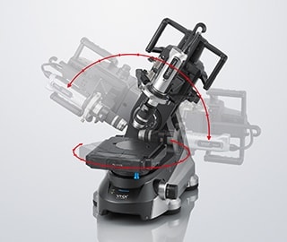
- Free-angle observation
-
With XYZ-axis control for easy adjustment of the field of view, the rotation axis, and the oblique axis, eucentric design ensures that the target stays centred in the field of view, even if the lens unit is tilted or rotated. Thanks to the long observation distance, important evidence can be observed at any angle with no contact. The depth composition function is also available during tilted observation.
- Quantified evidence evaluation with highly accurate 2D and 3D measurement and automatic analysis
-
The dimensions, area, volume, 3D shape, cross-section shape at an arbitrary location (profile), and surface roughness can be precisely measured with high-resolution 4K images in a non-destructive, non-contact manner. A wide range of automatic analysis functions, such as automatic area measurement/count, is also available, enabling quantitative analysis.
- Time reduction with automatic report creation
-
Excel can be installed directly on the VHX Series. Reports can be automatically created by outputting captured images and analysis results in arbitrary layouts using templates. This significantly reduces the time required for report creation, reducing the workload.

- 4K image capturing with hand-held observation using a microscope carried to the scene
-
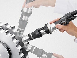
Usually, almost all work in forensics is performed in laboratories. However, in the case of buildings and for other such evidence that cannot be collected or moved, on-site, non-distractive and non-contact examination and inspection need to be performed. The VHX Series can capture on-site, high-resolution 4K images with hand-held observation.
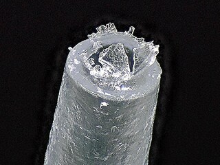
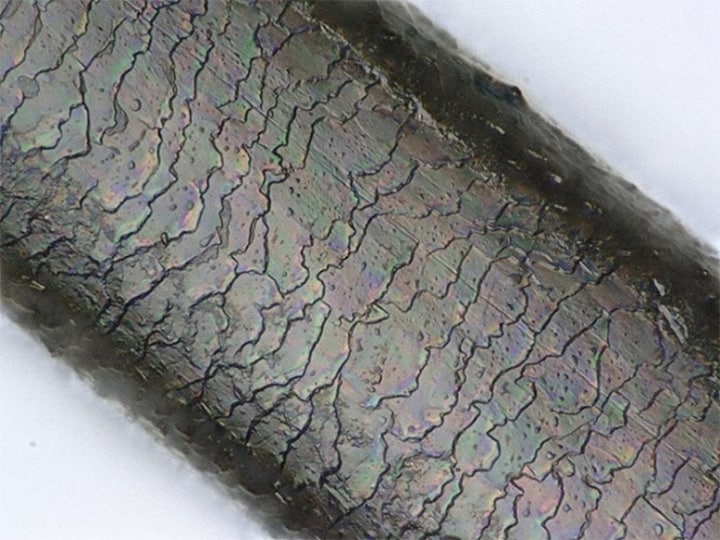
High-magnification observation of hair
Hair collected at scenes is important evidence for identifying perpetrators and people involved in incidents.
The VHX Series 4K digital microscope has a large depth of field, enabling observation with 4K images clearly focused on hair cuticles even at high magnifications.
This microscope is also equipped with a motorised revolver and a seamless zoom function, enabling automatic switching of the magnification from 20x to 6000x with no lens replacement. The magnification can be switched easily and quickly with a mouse or handheld controller while watching the screen.
In addition to hair, various types of evidence, such as skin, can be observed at high magnifications with higher sophistication and efficiency.
The VHX Series can precisely measure 2D and 3D dimensions using observation images. As such, the outer diameter and cross-section shape (profile) of hair can be measured in a non-destructive and non-contact manner, enabling speedy quantitative comparison and identification as well as analysis of various marks.

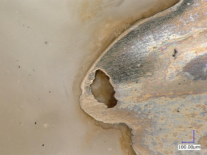
Observation of lustrous evidence such as metal dental crowns
Evidence can have various surface conditions. Lustrous evidence having curved surfaces—such as metal dental crowns, in particular—can diffuse reflected light, causing glare. This makes it difficult to determine lighting conditions, requiring a lot of time and effort.
The VHX Series 4K digital microscope can simplify lighting condition determination, enabling quick observation. This microscope is equipped with the Multi-lighting function, which automatically captures multiple images with omnidirectional lighting at the press of a button. The observation can be started simply by selecting the image from the multiple captured images that is visually most suitable for the purpose. This function greatly reduces the time required for condition determination.
Images other than that used for observation are also automatically stored and thus can be used again. Hence, there is no need to place the important evidence on the stage again to determine conditions even when the evidence needs to be observed under different conditions.
Additionally, by simply selecting a past image, the lighting conditions and settings used for capturing that image can be reproduced. Therefore, multiple samples of the same type of evidence can be observed under the same conditions, which is useful for quantitative examination.
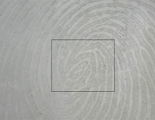
Capturing of fingerprints with high-resolution 4K images
Fingerprints are important marks for identifying people who have handled the evidence. However, it may be difficult to capture clear magnified images if the collected fingerprints have low contrast on the background.
The VHX Series 4K digital microscope simplifies the operations for light condition determination and other settings, enabling easy capturing of clear images even at high magnifications.
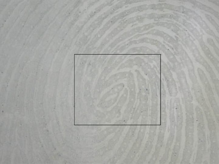
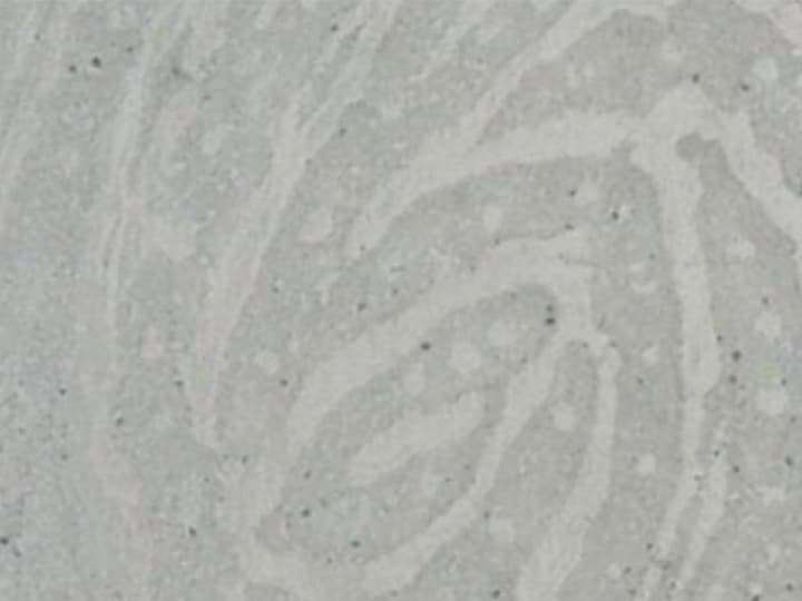
Fingerprints may have low contrast on the background during ordinary observation. This product is equipped with the High Dynamic Range (HDR) function, which captures multiple images at varying shutter speeds, generating an image with high colour gradation. This function allows 4K images to be captured with high resolution and high contrast.
High-magnification observation of fibres
To identify fibres, it is necessary to clearly capture high-magnification images of tissues having three-dimensional structures.
The VHX Series 4K digital microscope supports various lighting conditions with a single unit. For example, it is possible to clearly capture details such as differences in fibre glossiness and tissue structure by using coaxial lighting to receive specularly reflected light.
Additionally, with a large depth of field, the entirety of a three-dimensional tissue can be brought into focus even at high magnifications, which enables the capturing of 4K images suitable for identification.
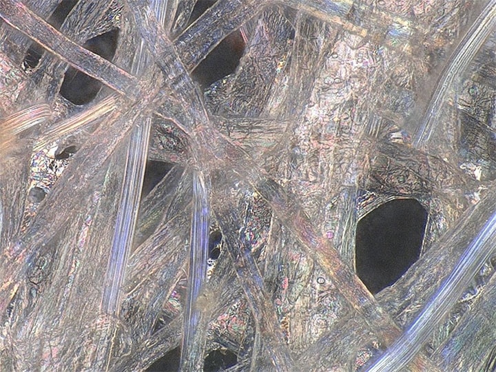
Image capturing of pollens in filters
Filters used for air cleaners and air conditioners have complex, three-dimensional fibre tissues. Optical microscopes cannot capture filter fibres and pollens adhering to these fibres simultaneously due to an insufficient depth of field that allows only a part of the field of view to be brought into focus.
The VHX Series 4K digital microscope is equipped with the depth composition function, which composes an image by capturing multiple images having different focus positions. Images fully in focus throughout the field of view can be captured even at high magnifications. With this function, it is possible to capture images focused on both the filter fibres having depth and the pollens adhering to these fibres.
Fully focused clear images obtained via depth composition help in identifying adhering substances in a non-destructive, non-contact manner even under difficult conditions like in fibre tissues of fabrics such as clothing and bedding.
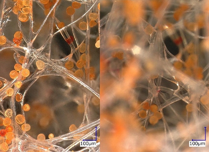
Additionally, the VHX Series can automatically perform highly accurate automatic area measurement/counting in the area specified by the operator on the image, enabling quick quantitative analysis of adhering substances.
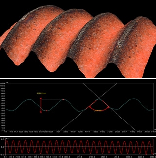
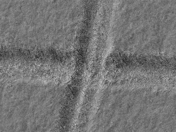
Handwriting analysis using high colour gradation images and colour map images
For handwriting identification, it is necessary to observe the details of marks left on paper fibres. However, as irregularities generated by writing pressure on the surface of paper are very small, it is difficult to scrutinise them using magnifying glasses or optical microscopes.
The VHX Series 4K digital microscope is equipped with Optical Shadow Effect Mode, which captures observation images having high colour gradation at the press of a button. As such, observation and analysis of even fine textures, minute surface irregularities, and subtle scratches are possible.
This product can perform observation in atmosphere, without the vacuum required by SEMs or the corresponding preparation time, ensuring that evidence stays free of damage.
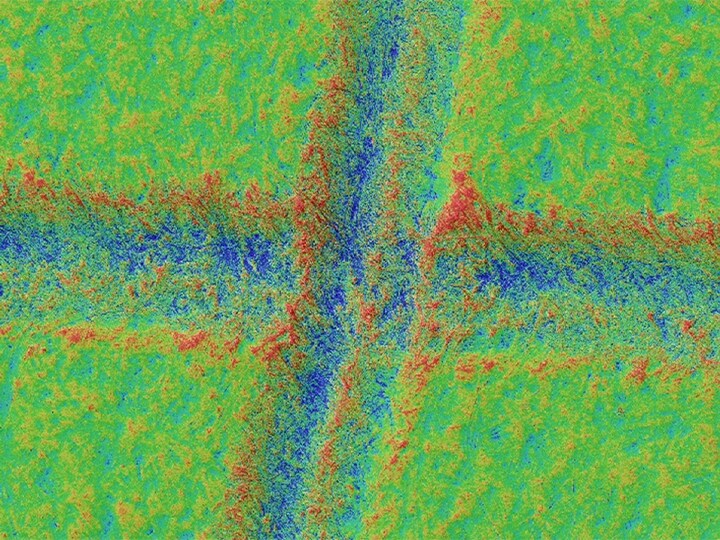
Images captured with Optical Shadow Effect Mode can also be displayed as a colour map. Subtle three-dimensional differences on a paper surface can be visualised with different colours, which is useful for analysis, comparison, and identification of patterns of writing pressure changes.
The following images are examples of the normal observation image, the Optical Shadow Effect Mode image, and the Optical Shadow Effect Mode colour map image of handwriting captured with the VHX Series.

A 4K Digital Microscope That Improves the Sophistication and Efficiency of Forensic Work Including Medical Jurisprudence
One of the major benefits that can be gained from using the VHX Series 4K digital microscope is usability that makes the high performance and various functions introduced above accessible quickly with simple operations.
The VHX Series can seamlessly perform the series of work from observation and analysis to automatic report creation with a single unit. This advantage increases the accuracy and efficiency of the large amount of daily forensic work, reducing the time required for examination and the workload of investigators.
Additionally, 4K images indicating the details of evidence can play a great role in showing the basis of examination results.
For additional info or inquiries about the VHX Series, click the buttons below.
